Respiratory System
Respiration is the process by which living organisms take in oxygen and release carbon dioxide. The human respiratory system, working in conjunction with the circulatory system, supplies oxygen to the body's cells, removing carbon dioxide in the process. The exchange of these gases occurs across cell membranes both in the lungs (external respiration) and in the body tissues (internal respiration). Breathing, or pulmonary ventilation, describes the process of inhaling and exhaling air. The human respiratory system consists of the respiratory tract and the lungs.
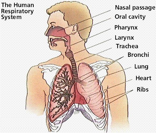
Respiratory tract
The respiratory tract cleans, warms, and moistens air during its trip to the lungs. The tract can be divided into an upper and a lower part. The upper part consists of the nose, nasal cavity, pharynx (throat), and larynx (voice box). The lower part consists of the trachea (windpipe), bronchi, and bronchial tree.
The nose has openings to the outside that allow air to enter. Hairs inside the nose trap dirt and keep it out of the respiratory tract. The external nose leads to a large cavity within the skull, the nasal cavity. This cavity is lined with mucous membrane and fine hairs called cilia. Mucus moistens the incoming air and traps dust. The cilia move pieces of the mucus with its trapped particles to the throat, where it is spit out or swallowed. Stomach acids destroy bacteria in swallowed mucus. Blood vessels in the nose and nasal cavity release heat and warm the entering air.
Air leaves the nasal cavity and enters the pharynx. From there it passes into the larynx, which is supported by a framework of cartilage (tough, white connective tissue). The larynx is covered by the epiglottis, a flap of elastic cartilage that moves up and down like a trap door. The epiglottis stays open during breathing, but closes during swallowing. This valve mechanism keeps solid particles (food) and liquids out of the trachea. If something other than air enters the trachea, it is expelled through automatic coughing.
Words to Know
Alveoli: Tiny air-filled sacs in the lungs where the exchange of oxygen and carbon dioxide occurs between the lungs and the bloodstream.
Bronchi: Two main branches of the trachea leading into the lungs.
Bronchial tree: Branching, air-conducting subdivisions of the bronchi in the lungs.
Diaphragm: Dome-shaped sheet of muscle located below the lungs separating the thoracic and abdominal cavities that contracts and expands to force air in and out of the lungs.
Epiglottis: Flap of elastic cartilage covering the larynx that allows air to pass through the trachea while keeping solid particles and liquids out.
Pleura: Membranous sac that envelops each lung and lines the thoracic cavity.
Air enters the trachea in the neck. Mucous membrane lines the trachea and C-shaped cartilage rings reinforce its walls. Elastic fibbers in the trachea walls allow the airways to expand and contract during breathing, while the cartilage rings prevent them from collapsing. The trachea divides behind the sternum (breastbone) to form a left and right branch, called bronchi (pronounced BRONG-key), each entering a lung.
The lungs
The lungs are two cone-shaped organs located in the chest or thoracic cavity. The heart separates them. The right lung is somewhat larger than the left. A sac, called the pleura, surrounds and protects the lungs. One layer of the pleura attaches to the wall of the thoracic cavity and the other layer encloses the lungs. A fluid between the two membrane layers reduces friction and allows smooth movement of the lungs during breathing.
The lungs are divided into lobes, each one of which receives its own bronchial branch. Inside the lungs, the bronchi subdivide repeatedly into smaller airways. Eventually they form tiny branches called terminal bronchioles. Terminal bronchioles have a diameter of about 0.02 inch (0.5 millimetres). This branching network within the lungs is called the bronchial tree.
The terminal bronchioles enter cup-shaped air sacs called alveoli (pronounced al-VEE-o-leye). The average person has a total of about 700 million gas-filled alveoli in the lungs. These provide an enormous surface area for gas exchange. A network of capillaries (tiny blood vessels) surrounds each alveoli. As blood passes through these vessels and air fills the alveoli, the exchange of gases takes place: oxygen passes from the alveoli into the capillaries while carbon dioxide passes from the capillaries into the alveoli.
This process—external respiration—causes the blood to leave the lungs laden with oxygen and cleared of carbon dioxide. When this blood reaches the cells of the body, internal respiration takes place. The oxygen diffuses or passes into the tissue fluid, and then into the cells. At the same time, carbon dioxide in the cells diffuses into the tissue fluid and then into the capillaries. The carbon dioxide-filled blood then returns to the lungs for another cycle.
Breathing
Breathing exchanges gases between the outside air and the alveoli of the lungs. Lung expansion is brought about by two important muscles, the diaphragm (pronounced DIE-a-fram) and the intercostal muscles. The diaphragm is a dome-shaped sheet of muscle located below the lungs that separates the thoracic and abdominal cavities. The intercostal muscles are located between the ribs.
Nerves from the brain send impulses to the diaphragm and intercostal muscles, stimulating them to contract or relax. When the diaphragm contracts, it moves down. The dome is flattened, and the size of the chest cavity is increased. When the intercostal muscles contract, the ribs move up and outward, which also increases the size of the chest cavity. By contracting, the diaphragm and intercostal muscles reduce the pressure inside the lungs relative to the pressure of the outside air. As a consequence, air rushes into the lungs during inhalation. During exhalation, the reverse occurs. The diaphragm relaxes and its dome curves up into the chest cavity, while the intercostal muscles relax and bring the ribs down and inward. The diminished size of the chest cavity increases the pressure in the lungs, thereby forcing air out.
A healthy adult breathes in and out about 12 times per minute, but this rate changes with exercise and other factors. Total lung capacity is about 12.5 pints (6 litters). Under normal circumstances, humans inhale and exhale about one pint (475 millilitres) of air in each cycle. Only about three-quarters of this air reaches the alveoli. The rest of the air remains in the respiratory tract. Regardless of the volume of air breathed in and out, the lungs always retain about 2.5 pints (1200 millilitres) of air. This residual air keeps the alveoli and bronchioles partially filled at all times.
Respiratory disorders
The respiratory system is open to airborne microorganisms and outside pollution. Some respiratory disorders are relatively mild and, unfortunately, very familiar. Excess mucus, coughing, and sneezing are all symptoms of the common cold, which is an inflammation of the mucous membrane lining the nose and nasal cavity. Viruses, bacteria, and allergens are among the causes of the common cold.
Since the respiratory lining is continuous, nasal cavity infections often spread. Laryngitis, an inflammation of the vocal cords, results in hoarseness and loss of voice. Viruses, irritating chemicals in the air, and overuse of the voice are causes of laryngitis.
Pneumonia, inflammation of the alveoli, is most commonly caused by bacteria and viruses. During a bout of pneumonia, the inflamed alveoli fill up with fluid and dead bacteria (pus). Breathing becomes difficult. Patients come down with fever, chills, and pain, coughing up phlegm and sometimes blood.
Sufferers of bronchitis, an inflammation of the bronchi, also cough up thick phlegm. There are two types of bronchitis, acute and chronic. Acute bronchitis can be a complication of a cold or flu. Bacteria, smoking, and air pollution can also cause acute bronchitis. This type of bronchitis clears up in a short time. Chronic bronchitis is a long-term illness that is mainly caused by air pollution and tobacco smoke. There is a persistent cough and congestion of the airways.
In emphysema, also caused by smoking, the walls of the alveoli disintegrate and the alveoli blend together. They form large air pockets from which air cannot escape. This cuts down the surface area for gas exchange. It becomes difficult for the patient to exhale. The extra work of exhaling over several years can cause the chest to enlarge and become barrel-shaped. The body is unable to repair the damage to the lungs, and the disease can lead to respiratory failure.
Asthma is a disorder of the nervous system. While the cause for the condition is unknown, it is known that allergies can trigger an asthma attack. Nerve messages cause extreme muscle spasms in the lungs that either narrow or close the bronchioles. A tightness is felt in the chest and breathing becomes difficult. Asthma attacks come and go in irregular patterns, and they vary in degree of severity.
Lung cancer is the leading cause of cancer death in men. It is the second leading cause of cancer death (after breast cancer) in women. Cigarette smoking is the main cause of lung cancer. Air pollution, radioactive minerals, and asbestos also cause lung cancer. The symptoms of the disease include a chronic cough from bronchitis, coughing up blood, shortness of breath, and chest pain. Lung cancer can spread in the lung area. Unchecked, it can spread to other parts of the body.
Advertise With Us
Latest Health News
- Carlton, AFL release statements on Elijah Hollands after mental health episode - afl.com.au
- AFL asks for Blues' report on Hollands health drama - ESPN
- Blues aware Hollands was 'struggling in the game' as AFL demands answers - Australian Broadcasting Corporation
- How Australia’s other half heals: all aboard the superyacht where rehab costs $600,000 a week - The Guardian
- Free RSV vaccine for older Australians - Australian Government Department of Health, Disability and Ageing